Dr. Yves Kayser - In-house X-ray spectroscopy

- Dr. Yves Kayser
- Project leader
- In-house X-ray spectroscopy
- Inorganic Spectroscopy
- +49 (0)208 306 - +49 (0)208 306 3694
- yves.kayser(at)cec.mpg.de
- Room: 512
Vita
| B.Sc. (Physics) | University of Fribourg, Switzerland (2005) |
| M.Sc. (Physics) | University of Fribourg, Switzerland (2007) |
| Ph.D. (Physics) | University of Fribourg, Switzerland (2011) |
| PostDoc | University of Fribourg, Switzerland (until 2012) Paul Scherrer Institut, Switzerland (until 2016) Physikalisch-Technische Bundesanstalt, Germany (until 2022) |
| Project leader | MPI CEC (since 2022) |
Publications
Full publications list | ORCID
Selected publications
- Wählisch, A., Wansleben, M., Weser, J., Stadelhoff, C., Holfelder, I., Kayser, Y. R., Beckhoff, B. (2023). Experimental determination of differential scattering coefficients for nickel by means of linearly polarized x-ray radiation (vol 60, 035001, 2023). Metrologia, 60(5): 059501, pp. 1-2. doi:10.1088/1681-7575/acee05.
- Hönicke, P., Kayser, Y. R., Soltwisch, V., Waehlish, A., Wauschkuhn, N., Scheerder, J. E., Fleischmann, C., Bogdanowicz, J., Charley, A.-L., Veloso, A., Loo, R., Mertens, H., Hikavyy, A., Siefke, T., Andrle, A., Gwalt, G., Siewert, F., Ciesielski, R., Beckhoff, B. (2023). Small target compatible dimensional and analytical metrology for semiconductor nanostructures using X-ray fluorescence techniques. In METROLOGY, INSPECTION, AND PROCESS CONTROL XXXVII. doi:10.1117/12.2657963.
- Wählisch, A., Wansleben, M., Weser, J., Stadelhoff, C., Holfelder, I., Kayser, Y. R., Beckhoff, B. (2023). Experimental determination of differential scattering coefficients for nickel by means of linearly polarized x-ray radiation. Metrologia, 60(3): 035001, pp. 1-13. doi:10.1088/1681-7575/acca87.
- Wach, A., Zou, X., Wojtaszek, K., Kayser, Y. R., Garlisi, C., Palmisano, G., Sa, J., Szlachetko, J. (2023). Towards understanding the TiO2 doping at the surface and bulk. X-Ray Spectrometry, (xx), 1-8. doi:10.1002/xrs.3363.
X-ray spectroscopy
In the Department of Inorganic Spectroscopy at the Max Planck Institute for Chemical Energy Conversion (MPI CEC) advanced high energy resolution X-ray spectroscopy is part of the analytical toolset used for studying fundamental chemical processes for energy storage in chemical molecules [1]. X-ray absorption spectroscopy (XAS) and X-ray emission spectroscopy (XES), which can be realized using synchrotron radiation, but also using laboratory-based stand-alone X-ray sources, allow probing the electronic and geometric structure of a catalytic material on an element selective basis. This knowledge contributes to a better understanding of the properties of catalytic materials and may provide clues towards their rational design.
The geometrical structure can be probed by means of extended X-ray absorption fine structure (EXAFS) with high sensitivity in terms of distance to and type of neighboring atoms and coordination number [2]. The electronic structure can be investigated from the X-ray absorption edge structure (commonly used acronyms are XANES for X-ray absorption near edge structure or NEXAFS for near edge X-ray absorption fine structure) or via XES by monitoring the energy differences between the different electronic levels involved in the monitored transitions. Transitions involving valence orbital are mostly of interest since their electron occupancy and energy impacts the possible formation of chemical bonds.
For 3d transition metals, which are attractive due to their abundancy, XANES is most commonly applied to obtain information about the local ligand field in the pre-edge region and the oxidation and spin state in the vicinity of the ionization threshold. XES measurements at the Kβ mainline offer sensitivity to the oxidation and/or metal spin state as well as the metal-ligand covalency [3], while monitoring the valence-to-core (VtC) provides information about ligand identity, electronic structure and metal-ligand bond length [4]. Note, that XANES and XES deliver not only complementary information by probing the unoccupied, respectively the occupied electronic density of states, but the use of both analytical techniques may enable a more unambiguous interpretation of the different high energy resolution spectra by disentangling the influence of different parameters on the spectral shape, e.g., ligand identity or coordination [5,6].
Currently, the named high energy resolution X-ray spectroscopies are most commonly applied at synchrotron radiation facilities where high brilliance in combination with energy tunability are offered. These advantage permit to realize advanced X-ray spectroscopy like resonant XES (RXES) or high resolution fluorescence detected XAS, but the experimental time that can be allocated at these large-scale facilities is limited. Energy-dispersive schemes allow, however, to realize XANES and EXAFS in transmission mode in stand-alone configurations, which do not depend on probing X-ray radiation being delivered by a large-scale facility. Non-resonant XES measurements can be likewise realized with stand-alone instrumentation since monochromatization of the photons used for excitation is not mandatory. Stand-alone instrumentation which is suitable for in-house use is highly advantageous for frequent and flexible access and thus for investigations requiring a quick response time, regular access or the handling of delicate samples. It should not be neglected that in-house facilities can also serve educational and training purposes, both in terms of experimental execution as well as in terms of data analysis.
In this view, a collaboration was established with the Analytical X-ray physics group of the Technical University of Berlin and the Berlin Laboratory for Innovative X-ray Technologies which pioneered the development of efficient stand-alone high energy resolution X-ray spectrometers. In particular the use of highly annealed pyrolithic graphite (HAPG) was pioneered. The difference between this material and pure crystals is its high integral reflectivity such that a larger fraction of photons can be diffracted and detected at the expense of the energy resolution achievable. The instrumentation realized allows for XANES or EXAFS experiments in transmission-mode in the soft to the hard X-ray energy range, respectively for XES in the tender to hard X-ray energy range. The different spectrometers mentioned are realized in the dispersive mode, meaning that within an energy range of interest all relevant photon energies are monitored simultaneously in a static configuration without moving any spectrometer components. This aspect is highly beneficial for realizing efficient experiments since the best use of the photons generated by the stand-alone X-ray source is being made.
Details on the three in-house X-ray spectrometers realized for laboratory-based high energy resolution X-ray spectroscopy investigations are as follows.
In the soft energy range a laser plasma source is used for generating a broad energy range X-ray spectrum in the energy range from about 200 eV up to 1 keV and at a repetition rate of 100 Hz, while a grating and CCD camera are used for the dispersion and position-sensitive detection of the generated X-rays. The basic concept is reported in [7] and the instrument after installation is displayed in Figure 1. In the experimental setup realized at the MPI CEC, two branches are connected to the laser plasma source. For each branch two gratings are in use, one where the X-rays transmitted through a sample are subject to measurement and one where a reference spectrum is being recorded (i.e., no sample in the beam path from the source to the grating). The two branches are designed to accommodate either solid or liquid sample environments (in cells or using a jet). Static and transient X-ray absorption spectroscopy using a pump laser in the wavelength range from 210 nm to 2600 nm can be realized on both branches. Given the pulse duration of the order of nanoseconds for the pump and probe laser, good time resolution can be expected for the transient spectrosopy experiments.

In the hard X-ray range, EXAFS experiments can be realized in transmission mode in a dispersive scheme using a spectrometer based on the von Hamos geometry. The accessible energy range is 4 keV to 15 keV. A description of the spectrometer concept is reported in [8]. HAPG crystal is used as the diffraction crystal and a hybrid pixel counting detector for the spatially resolved detection. The detector is oriented differently than in the conventional von Hamos geometry to allow for monitoring a broader energy range at a single position of the components and compensating for sagittal focusing errors from the diffraction crystal. Appropriate operation conditions of the X-ray tube exclude the detection of higher diffraction orders. The sample is positioned between the X-ray tube and the diffraction crystal. In future optional cryo-cooling will be made available for static experiments. Furthermore, routes for enabling in situ and operando conditions for investigating catalytic materials will be explored by implementing heating and gas flow options.
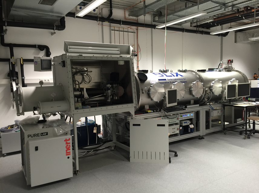
For XES experiments in the tender to hard X-ray range a full-cylinder von Hamos spectrometer based on a HAPG crystal and a CCD camera van be used in the energy range from 2.4 keV to 22 keV. The full cylinder concept ensures an enhanced solid angle of detection compared to the conventional von Hamos geometry which allows in conjunction with the inherent high integral reflectivity of the mosaic HAPG crystals and the liquid metal jet used for XES experiments with optimized excitation and detection conditions [9]. This in-vacuum spectrometer offers the option to perform experiments on cryo-cooled samples. Moreover, the spectrometer is directly connected to an Ar filled glove-box for on-site sample preparation in an inert atmosphere just prior to the XES measurements (Figure 2).

As a complementary instrument to the EXAFS and XES spectrometers described above in terms of sample environments that can be implemented in the X-ray spectroscopy investigations and analytical capabilities offered, a commercial spectrometer from easyXAFS for XAS (XANES and EXAFS) and XES experiments in the X-ray energy range form 5 keV to 23 keV is available [10]. The easyXAFS spectrometer is combined with a conventional transmission-type X-ray tube and is based on the Rowland circle geometry: exchangeable spherically curved perfect crystals are being used for the Bragg diffraction of the X-rays and an energy-dispersive detector for their detection. In contrast to the previous spectrometers mentioned, measurements need to be realized in a scanning-type fashion. Current developments on the sample environment include sample heating, gas flow and the application of an electric potential.
In summary, the in-house X-ray spectroscopy instrumentation aims at supporting the understanding on an atomic level of the geometric and electronic structure of catalytic materials and in the future possibly of the changes in structure occurring during catalytic reactions. To this end, future instrumentation development directions include sample environments such that investigations under in situ and operando conditions become accessible. Considering time-resolved experiments, the accessible time scale can range from minutes to hours such that X-ray damage mechanisms, long-term stability and deactivation processes become accessible. For the best time resolution and sensitivity, advanced X-ray spectroscopy experiments at synchrotron radiation facilities remain the method of choice, for which in-house X-ray spectroscopy investigations can be an essential tool for the preparation and optimization of experimental conditions and estimation of the achievable sensitivity and discrimination capabilities. Thus, the realized in-house high energy resolution X-ray spectroscopy instrumentation can present a valuable tool towards linking catalytic activity to changes in the electronic and geometric structure during reactions and thus in obtaining clues about the catalyst working mechanisms.
References
[1] G. E. Cutsail III and S. DeBeer, Challenges and Opportunities for Applications of Advanced X‑ray Spectroscopy in Catalysis Research, ACS Catal. 2022, 12, 5864−5886.
[2] J. Timoshenko and B. Roldan Cuenya, In Situ/Operando Electrocatalyst Characterization by X‑ray Absorption Spectroscopy, Chem. Rev. 2021, 121, 882–961.
[3] C. J. Pollock, M. U. Delgado-Jaime, M. Atanasov, F. Neese and S. DeBeer, Kβ Mainline X ray Emission Spectroscopy as an Experimental Probe of Metal-Ligand Covalency, J. Am. Chem. Soc. 2014, 136, 9453−9463.
[4] N. Lee, T. Petrenko, U. Bergmann, F. Neese and S. DeBeer, Probing Valence Orbital Composition with Iron Kβ X-ray Emission Spectroscopy, J. Am. Chem. Soc. 2010, 132, 9715–9727.
[5] L. Decamps, D. B. Rice, and S. DeBeer, An Fe6C Core in All Nitrogenase Cofactors, Angew. Chem. Int. Ed. 2022, 61, e202209190.
[6] Z. Mathe, D. A. Pantazis, H. Beom Lee, R. Gnewkow, B. E. Van Kuiken, T. Agapie, and S. DeBeer, Calcium Valence-to-Core X-ray Emission Spectroscopy: A Sensitive Probe of Oxo Protonation in Structural Models of the Oxygen-Evolving Complex, Inorg. Chem. 2019, 58, 23, 16292–16301.
[7] A. Jonas, H. Stiel, L. Glöggler, D. Dahm, K. Dammer, B. Kanngießer and I. Mantouvalou, Towards Poisson noise limited optical pump soft X-ray probe NEXAFS spectroscopy using a laser-produced plasma source, Opt. Express 2019, 27, 36524-36537.
[8] C. Schlesiger, S. Praetz, R. Gnewkow, W. Malzer and B. Kanngießer, Recent progress in the performance of HAPG based laboratory EXAFS and XANES spectrometers, J. Anal. At. Spectrom. 2020, 35, 2298-2304.
[9] W. Malzer, D. Grötzsch, R. Gnewkow, C. Schlesiger, F. Kowalewski, B. van Kuiken, S. DeBeer and B. Kanngießer, A laboratory spectrometer for high throughput X-ray emission spectroscopy in catalysis research, Rev. Sci. Instrum. 2018, 89, 113111.
[10] E. Jahrmann, W. M. Holden, A. S. Ditter, D. R. Mortensen, G. T. Seidler, T. T. Fister, S. A. Kozimor, L. F. J. Piper, J. Rana, N. C. Hyatt, and M. C. Stennett, An improved laboratory-based x-ray absorption fine structure and x-ray emission spectrometer for analytical applications in materials chemistry research, Rev. Sci. Instrum. 2019, 90, 024106.
